


Pleural Fluid Analysis
Dr Babina Thangjam *
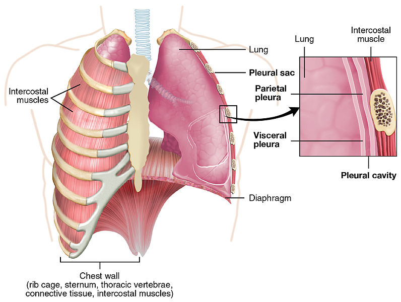
The Lung Pleurea : Illustration from Anatomy & Physiology, Connexions Web site. http://cnx.org/content/col11496/1.6/, Jun 19, 2013.
Pix Courtesy: Wikipedia/OpenStax College
Pleural fluid is the fluid that is normally present in the pleural space. Pleural space is the space between the lining of the outside of the lungs (pleura) and the wall of the chest. This serves as a lubricant for the movement of the lungs during inhalation and exhalation. When excess fluid collects in the pleural space, the condition is called pleural effusion.
A variety of conditions and diseases can cause inflammation of the pleurae (pleuritis) and/or excessive accumulation of pleural fluid (pleural effusion). Pleural fluid analysis is a group of tests that evaluate this fluid to determine the cause of the increased fluid.
TYPES OF PLEURAL FLUID
The two main reasons for the accumulation of pleural fluid in the pleural space are:
o When an imbalance occurs between the pressure of the liquid within blood vessels (which drives fluid out of blood vessels) and the amount of protein in blood (which keeps fluid in blood vessels), excess fluid gets collected in the pleural cavity. This type of fluid is called a TRANSUDATE. This type of fluid usually involves both lungs.
o When there is an injury to or inflammation of the pleurae, extra fluid gets accumulated in the pleural space. This accumulated fluid is called an EXUDATE. It usually involves one lung
Differentiation between the types of fluid through tests is important because it helps diagnose the specific disease or condition. Once the type of the fluid identified, additional tests may be performed to further pinpoint the disease or condition causing pleuritis and/or pleural effusion.
HOW IS THE SAMPLE COLLECTED?
A sample for pleural fluid is collected by a doctor with a syringe and needle using a procedure called thoracentesis.
PREPARATION NEEDED FOR THE TEST
No special preparation is needed for this test. However, a chest X-ray is usually done before the test.
HOW IS THE TEST USED?
An initial set of tests is used to differentiate between the two types of fluid that may be produced.
When the fluid is confirmed to be transudate fluid, then usually no more tests on the fluid are necessary. Transudates are most often caused by either congestive heart failure or cirrhosis. However, sometimes fluid analysis are done on transudates which include:
o Physical appearance.
o Protein, albumin.
o Cell count.
Exudates are associated with a variety of conditions and diseases that include:
o Infectious diseases that are caused by viruses, bacteria or fungi.
o Bleeding disorders, pulmonary embolism, or trauma.
o Inflammatory conditions such as lung diseases and chronic lung inflammation, or autoimmune disorders.
o Malignancies.
o Other conditions like cardiac bypass surgery, heart or lung transplantation, pancreatitis, or intra-abdominal abscesses.
Additional testing on exudate fluid may include:
o Microscopic examination - Normal pleural fluid has small numbers of white blood cells (WBCs) but no red blood cells (RBCs) or microorganisms. Laboratories may examine the pleural fluid on a slide. The slide is treated with a special stain and evaluated for the different kinds of cells that may be present.
o Gram Stain For direct observation of bacteria or fungi under a microscope. There should be no organisms present in the normal pleural fluid.
o Bacterial culture and susceptibility testing They are done to detect any microorganisms that may be present in the pleural fluid and to guide antimicrobial therapy.
o Tests for infectious diseases, such as tests for viruses, mycobacteria (AFB smear and culture), and parasites. Tuberculous pleural effusion is one of the most common forms of extrapulmonary tuberculosis (TB). The diagnosis of a tuberculous pleural effusion requires a positive culture (from pleural fluid or pleural tissue) or the presence of granulomas in the pleura.
o Pleural fluid glucose, lactate, amylase, triglyceride and/or tumour markers.
WHEN IS THE TEST DONE?
Pleural fluid analysis may be done when a doctor suspects that a person has a condition or disease that is causing pleuritis and/or pleural effusion. A patient with pleural effusion may have the following signs and symptoms:
o Chest pain that worsens with deep breathing.
o Coughing, fever and chills.
o Difficulty breathing, shortness of breath.
* Dr Babina Thangjam, MD wrote this article for The Sangai Express
The writer is Consultant Pathologist, BABINA Diagnostics, Imphal.
This article was posted on August 14 , 2015.
* Comments posted by users in this discussion thread and other parts of this site are opinions of the individuals posting them (whose user ID is displayed alongside) and not the views of e-pao.net. We strongly recommend that users exercise responsibility, sensitivity and caution over language while writing your opinions which will be seen and read by other users. Please read a complete Guideline on using comments on this website.


