


What if sudden deaths of piglets or adult pigs are due to SWINE FEVER disease?
- Part 1 -
Dr. Shongsir Warson Monsang / Dr. Rajkumari Mandakini *
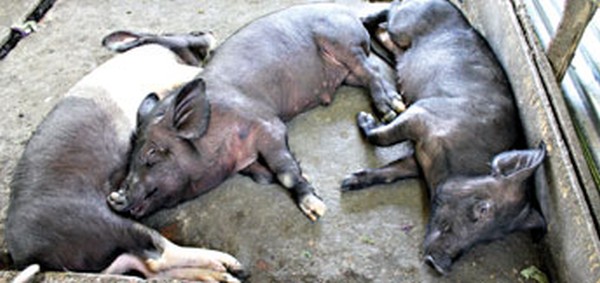
File picture of ailing pigs at a piggery :: Pix - TSE
Reports highlighted in the local newspapers (2013) regarding the sudden death of piglets or adult pigs caused a panic for all the people residing in Manipur for one or the other reason as it posed a serious human health concern. Manipur is a state, well known for its consumption of meat inclusive of white as well as red meat. Amongst the marketed meat, pork is considered one of the people's choices because of its palatability and back fat thickness. Consumption of meat is not an issue unless found unfit for any disease which may affect the health of the consuming individual or the family.
Scientifically proven, there are many diseases of the animals which can be transmitted to the human population called "Zoonotic" disease. These diseases may enter into human beings through several routes which may even lead to the loss of life in the absence of prompt diagnosis and preventive measures.
What if the results of the laboratory conferred to positive swine fever? This is the very question which would hit the head of every intellectual and literate people because the disease is economically important to the state so far. Taking the interests of the welfare of our people, this article is presented in a very simple and understandable form so that one person from each family can be instrumental in spreading the awareness and help in undertaking precautionary programmes as and when it is required.
Swine Fever is a notifiable disease of pigs which is considered a highly infectious as well as contagious disease caused by a virus belonging to genus Pestivirus of the Flaviviridae family. The natural reservoirs of classical swine fever are pigs and wild boar and all feral and wild pigs including European wild boar are found to be most susceptible. The severity of this disease varies with the strain of the virus, the age of the pig, and the immune status of the herd. Acute infections which are caused by highly virulent isolates have a high mortality rate and are likely to be diagnosed rapidly. However, infections with less virulent isolates can be more difficult to recognize, particularly in older pigs.
Transmissions: Pigs can become infected with the virus after eating contaminated foods, which mostly occurs from feeding of undercooked or uncooked garbage or meat products. These pigs are exposed to infected pigs or objects (fomites) contaminated by infected pigs, example equipments, boots, clothing, buckets, etc. The viruses are shed in the blood, saliva, nasal discharge, secretions and excretions (urine, faeces, etc.) or tissues of infected pigs. The virus can also be spread by infected pigs in semen during breeding or across placenta during gestation; giving birth to persistently infected piglets. Airborne transmissions or spread by aerosol vectors, example flies usually seem possible over short distances.
CSFV is moderately fragile in the environment and can survive for 3 days at 50ºC and 7 to 15 days at 37ºC. CSFV can remain infectious for nearly three months in refrigerated meat and for more than four years in frozen meat. In this proteinaceous environment, this virus does not appear to be inactivated by smoking or salt curing.
Clinical signs: The symptoms and severity of the disease varies with the strain of the virus, age and susceptibility of the piglets. The more virulent strains caused acute form of the disease, while the less virulent strain results in chronic, mild or asymptomatic infections in a high percentage of the animal.
Acute form: Considered as the most severe form which occurs rapidly (within 2-15 days) resulting in variable rates of illness and death. Common symptoms include a high fever (105-107°F), huddling, weakness, drowsiness, anorexia, intermittent constipation followed by diarrhoea and conjunctivitis. Pigs may exhibit an unsteady, staggering gait which progress to posterior paresis.
The abdomen, inner thighs, ears and tail may develop a purple cyanotic discoloration. Haemorrhages can also occur in the skin. Severe leukopenia usually occurs soon after the disease onset and convulsions may be seen in the terminal stages. Pigs with acute classical swine fever often die within 1-3 weeks. The mortality rate can be as high as 90 per cent. Pneumonia is a common post-mortem finding and 'button ulcers' may be present in the intestines.
Subacute form: The subacute form is similar to acute classical swine fever; however, the symptoms are less severe, and the fever may persist for two to three weeks. Some pigs with subacute classical swine fever may survive; others die within a month.
Chronic form: Occurs ever a long period of time (2-4 weeks) and may affect only a few animals. In the initial stages, chronic disease can resemble acute or subacute disease, with anorexia, depression, elevated temperatures, leukopenia, and periods of constipation and diarrhoea. Affected pigs usually improve after several weeks; however, after a period where they appear relatively normal, they develop recurrent symptoms that may include intermittent fever, anorexia, periods of constipation or diarrhoea, wasting or stunted growth, alopecia and skin lesions. Immunosuppression may lead to concurrent infections. Affected pigs may survive for 1-3 months, but chronic infections are always fatal.
Post Mortem Lesions: The lesions of classical swine fever are highly variable.
In acute disease, the most common lesion is haemorrhage. The skin may be discoloured purple and the lymph nodes may be swollen and hemorrhagic. Petechial or ecchymotic haemorrhages can often be seen on serosal and mucosal surfaces, particularly on the kidney, urinary bladder, epicardium, larynx, trachea, intestines, subcutaneous tissues, and spleen. Strawcolored fluid may be found in the peritoneal and thoracic cavities and the pericardial sac. Splenic infarcts are occasionally seen. The lungs may be congested and hemorrhagic. In some acute cases, lesions may be absent or inconspicuous.
The lesions of chronic disease are less severe and may be complicated by secondary infections. In addition, necrotic foci or "button" ulcers may be found in the intestinal mucosa, epiglottis and larynx. In growing pigs that have survived for more than a month, bone lesions can also occur at the costochondral junction of the ribs and the growth plates of the long bones.
In congenitally infected piglets, common lesions include cerebellar hypoplasia, thymic atrophy, ascites, and deformities of the head and legs. Edema and petechial hemorrhages may be seen in the skin and internal organs.
To be continued....
* Dr. Shongsir Warson Monsang, (Ph.D), Surgery / Dr. Rajkumari Mandakini, (Ph.D Scholar), Bacteriology wrote this article for e-pao.net
Dr. Shongsir Warson Monsang can be contacted at warsonmonsang(at)gmail(dot)com
This article was posted on February 06, 2014.
* Comments posted by users in this discussion thread and other parts of this site are opinions of the individuals posting them (whose user ID is displayed alongside) and not the views of e-pao.net. We strongly recommend that users exercise responsibility, sensitivity and caution over language while writing your opinions which will be seen and read by other users. Please read a complete Guideline on using comments on this website.


