


Ultrasound
Dr Momocha Thangjam *
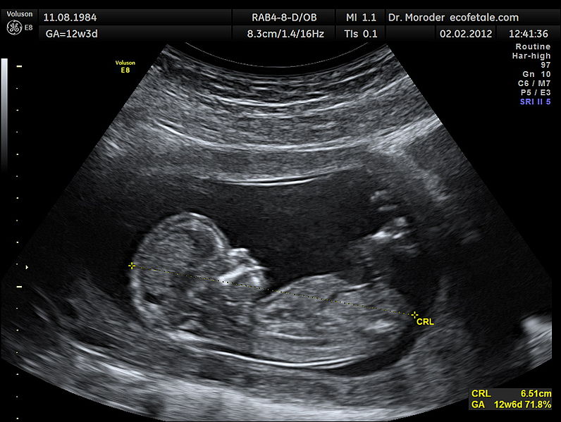
Ultrasound image of the foetus at 12 weeks of pregnancy in a sagittal scan. Measurements of fetal Crown Rump Lenght (CRL).
Pix - wikipedia/Moroder
Diagnostic ultrasound, also called sonography is an imaging technique that uses high-frequency sound waves to produce relatively precise images of structures within the body. The technique is similar to the echo-location used by bats, whales and dolphins, as well as SONAR used by submarines.The images produced during an ultrasound examination provide valuable information for diagnosing and treating a variety of diseases and conditions.
Ultrasound was first used for clinical purposes in 1956 in Glasgow. By the end of the 20th century, ultrasound imaging has become routine in maternity clinics throughout the world.
USES OF ULTRASOUND
Ultrasound may be used to:
o Assess a foetal growth and development during pregnancy.
o Diagnose diseases and cancer of liver,pancreas, and other abdominal organs.
o Detect stones and cancer of kidneys and gall bladder.
o Diagnose acute conditions like appendicitis.
o Evaluate a breast lump, thyroid gland or eye problems.
o Diagnose diseases of prostate and testes in males and uterus and ovaries in females.
o Evaluate abnormalities of the muscles.
o Detect fluid in lungs or inside abdomen.
o Evaluate brain in newborns.
o Guide a needle for FNAC/biopsy.
LIMITATIONS
Although ultrasound is a valuable tool, it does have its limitations. Sound does not travel well through air or bone, so ultrasound is not effective at imaging parts of the body that have gas in them or that are obscured by bone. Rather than using ultrasound to view these areas, other imaging tests, such as CT/ MRI scans or X-rays are preferred.
PREPARATIONS
o Most ultrasound exams require no preparation.
o Gallbladder examination requiresfasting for up to six hours before the examination.
o A pelvic ultrasound requiresholding of urine before the examination to ensure filling of bladder to allow better visualisation of the uterus, ovaries or prostate.
PROCEDURE
During an ultrasound examination, the patient lies on an examination table. A small amount of gel is applied to the skin. The gel helps eliminate the formation of air pockets between the ultrasound and the body. During the exam, the radiologist/sonologist presses a small hand-held device (transducer) against the skin over the area of the body being examined. Though the majority of ultrasound exams are performed with a transducer on the skin, some ultrasounds are done inside the body with the help of probes inserted through natural openings of body such as Transrectal, Transvaginal and Transesophageal ultrasound.
Ultrasound is a painless procedure. However, patient may experience some mild discomfort in transrectal/transvaginal scans. A typical ultrasound examination takes a few minutes. The radiologist finally analyses the images and gives the report.
BENEFITS
Ultrasound is one of the most widely used imaging modality. It is safe, relatively inexpensive and widely available. It can be used to diagnose a wide range of diseases.
RISKS
Diagnostic ultrasound is a safe procedure that uses low-power sound waves. There are no direct risks from an ultrasound exam.
RECENT ADVANCE
Latest developments in Ultrasound include 3D/4D Real time volume imaging for abdominal organs and foetus, Intra-vascular ultrasound, Elastography for breast lumps, use of contrastagents for better detection of cancers etc.
PCPNDT Act:
The Pre-Conception and Pre-Natal Diagnostic Techniques (Prohibition Of Sex Selection) Act is federal legislation enacted by the Parliament of India to stop female foeticides and arrest the declining sex ratio in India. The main purpose of enacting the Act is to ban the use of sex selection techniques before or after conception and prevent the misuse of prenatal diagnostic technique for sex selective abortion.
Sex detection is punishable with imprisonment for a term which may extend from three to five years and with fine which may extend from ten thousand to one lakh rupees.
Punishment is both for the doctor who does sex determination and the parents/relatives who seek sex determination. Sex determinationis allowed on stringent medical grounds i.e. when chromosomal/genetic abnormalitiesis suspected in the foetus or a hereditary disease runs in the family (e.g.sex-linked disorders).
o Dr Momocha Thangjam, DMDRD, DNB wrote this article for The Sangai Express
The writer is Consultant Radiologist, BABINA Diagnostics, Imphal
This article was posted on April 16, 2014.
* Comments posted by users in this discussion thread and other parts of this site are opinions of the individuals posting them (whose user ID is displayed alongside) and not the views of e-pao.net. We strongly recommend that users exercise responsibility, sensitivity and caution over language while writing your opinions which will be seen and read by other users. Please read a complete Guideline on using comments on this website.


